X-Rays – Production, Properties, Wavelength and Uses
Roentgen’s discovery of X-rays
Roentgen’s discovery of X-rays :- In 1895, the German scientist Rontgen observed that when a fast moving cathode ray collides with a piece of metal having a higher atomic mass and a higher melting point.
A new type of ray is produced which is not visible by the eye. but in plate of photography It acts the same way as light rays do.
He named these unknown and invisible rays as X-Rays. In the name of roentgen or röntgen, these rays are also called Rontgen Rays. Professor Röntgen was awarded the noble prize of physics in 1901 for this important discovery.
Production of X Rays by Coolidge Tube
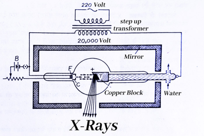
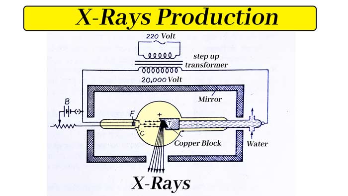
Nowadays Coolidge tubes are used to make X-rays. It was created in 1913 by Dr. Coolidge. It consists of a spherical bulb made of hard glass. Inside the glass there is a high vacuum of about 10-6 cm (Mercury). It has two drains connected.
A tube has a filament (F) of tungsten in which current is passed through a battery (B). When the filament is heated, electrons start coming out of it, due to thermic effect, whose number depends on the temperature of the filament.
There is a cylinder (C) of molybdenum around the filament which is kept at negative potential relative to the filament. Due to this the electrons coming out of the filament get concentrated in the form of a fine beam.
There is a block of copper just in front of the filament F whose plane is inclined at 45° to the path of the electron beam. A piece of metal with high melting point and high atomic mass like tungsten or molybdenum (T) is deposited on this plane.
It is called Anti-Cathode or Target. The copper block is located at the end of a hollow copper tube in which a stream of cold water is passed. The entire tubule is surrounded by a lead shell.

HowX-Ray is produced
working : When an alternating potential difference of about 20000 volts is applied between filament (F) and target (T) by a high-voltage transformer. The electrons coming out of the filament hit the target with a very high velocity and X-Rays start coming out.
The target (T) becomes very hot due to the constant collision of electrons.
In fact, only 2% of the energy of electrons is used to produce x rays, the remaining part gets converted into heat. If there is no arrangement to remove this heat, the target may melt.
For this reason, it is kept cool by flowing water in a copper tube around the target.
Importance of Biomolecules in Life || What are the 4 main biomolecules?
Resonance effect or mesomeric effect || What is resonance effect with example?
Valency of Elements || How to Find Valency || What is the Valency of the atom?
Glucose Structure: Physical and chemical properties, Glucose Chemical Reaction
Introduction of Inductive-Effect || How does Inductive Effect Work?
IUPAC Name : How to find the IUPAC name of compounds.
In the half cycle of alternating potential difference, when the target (T) is positive with respect to the filament (F), then the electron gets attracted and hits the target.
In the remaining half cycle, the target becomes negative with respect to the filament, due to which the electrons are repelled. In this way the electrons hit the target only in half the cycle and the tube itself acts like a rectifier.
For the generation of x rays it is necessary that the electrons from the filament do not collide with the atoms of the gas of the tube before reaching the target. That is why a high vacuum is maintained in the tube.
If this is not the case, the electron will lose its energy by ionizing the gas and the generated positive ions will collide with filament and damage the filament.
Control Over Intensity and Penetration of X-Rays
Soft and Hard X-Rays
In generating X-rays, their two properties have to be taken care of: Intensity and Penetration
Control over Intensity: The intensity of x-rays is called their rate of production i.e. the number of x-rays produced per second. Their value is directly proportional to the number of electrons producing x rays.
If the number of electrons hitting the target per second is increased, then the number of X rays produced will also increase i.e. X rays of more intensity will be produced.
To increase the intensity of X rays received from the Coolidge tube, the current flowing in the filament is increased. This increases the number of electrons per second from the filament, that is, the number of electrons hitting the target per second. As a result, the intensity of the generated X rays increases.
Control over Penetration: The penetrability of X rays depends on their wavelength. Rays with larger wavelength (approximately 4Å) have less frequency (v = c/λ), so their energy is also less.
When the energy (hv) is less, their penetrating power is less and they are called soft or less penetrating x rays.
These rays can pass through only thin sheets of matter. In contrast, rays with shorter wavelength (about 1Å) have higher penetrating power and are called hard or highly penetrating X rays.
The wavelength of X-Rays depends on the kinetic energy of the electrons producing them and this kinetic energy depends on the potential difference between the filament (F) and the target (T) inside the tube.
As this potential difference is increased, the wavelength of the X rays produced in the tube becomes smaller, that is, the penetrating power increases.
Therefore, the power of X rays can be increased by increasing the potential difference applied at the ends of the tube and it can be reduced by decreasing.
The doctor applies a potential difference of about 60000 volts to take X-ray of the patient’s body. These X rays cross the flesh.
But not the bones. If a potential difference of about 10 lac volts is applied, the rays will cross even thick iron sheets.
In this way the intensity and penetration of the rays received from the coolidge tube can be controlled independently.
Spectrum of X-Rays
A continuous spectrum of X-Rays of Coolidge Tube is obtained. This means that these rays consist of rays of many wavelengths, whose intensities merge with each other in such a way that no definite separating line can be drawn between the two wavelengths.
There is a lowest limit of wavelength λmin for X rays. The X rays coming out of the tube do not contain X rays of wavelength smaller than λmin. The value of λmin depends on the voltage V of the tube.
The higher the voltage, the lower the value of λmin.
Explain: The emission of X-rays due to collision of electrons on the target of the tube and the lowest wavelength limit can be explained by quantum theory. According to this theory, X rays are emitted in the form of small bundles of energy which are called Photons.

The amount of energy associated with each photon is hv, where v, is the frequency of X radiation and h is the plank constant.
When V voltage is applied to the X ray tube, the energy of the electrons coming out of the filament to reach the target will be eV, where e is the charge of the electron.
When such an electron collides with an atom of the target, it loses some of its energy. This lost energy is obtained in the form of photon of X ray whose energy is hv.
It is clear that the value of hv will be less than eV.
Calculation of the minimum limit of the Wavelength:
The electron collides with a number of atoms of the target before reaching a steady state, decreasing the energy of the photons being emitted.
In addition, if the electron gives all its energy eV to collide with only one atom, then a photon of maximum energy hvmax is emitted.
Thus, the X radiation emitted from the target will have a continuous range of frequencies and the maximum frequency will be vmax.

The minimum wavelength corresponding to the maximum frequency is λmin.
Where c is the light speed. Substituting the value of vmax from equation (i) in equation (ii)
Hence, the lowest wavelength limit (λmin) is inversely proportional to the accelerating potential (V).
Solving the above formula by keeping h = 6.6 x 10⁻³⁴ joule second, c = 3.0 x 10⁸ meter/second and e = 1.6 x 10⁻¹⁹ coulomb
In this formula the value of accelerator potential (V) is in volt.
Properties of X-Rays
(i) X rays are electromagnetic rays like light rays. All the major properties of the light rays are found in them:
a) It is not visible.
b) It moves in a straight line.
c) It moves with the speed of light.
d) They have reflection, refraction, interference, diffraction and polarisation.
(iii) X-Rays are not deflected in electric and magnetic fields.
(iv) These rays give glow when they fall on the fluorescent materials. For example, it illuminates the postulate of berium platino cyanide. Using these properties, doctors examine the internal organs of the patient.
(v) The gas through which the X-rays fall, ionizes it.
(vi) These rays penetrate to different depths in different materials like wood, cardboard, thin sheet of metal, meat, etc. Their capacity depends on their wavelength. It does not pass through heavy objects and bones. If these substances are kept in their path, then their shadow is formed.
(vii) It darkens the photographic plate by chemical reaction.
(viii) It is very active. If they are put on a zinc plate, then electrons can be removed from it.
(ix) If these rays are put on the human body for a long time then it is fatal.
(x) When x rays are put on a metal plate, some part goes across and the remaining part gets converted into different types of secondary radiations.
Applications of X-Rays
Till date no other scientific investigation has done as many uses of X rays in various fields.

In surgery: X rays easily penetrate through low density materials like wood, paper, meat and blood, but cannot pass through high density objects like bone, iron.
Using this quality, rotten or broken bones, sunken bullets or stones and lung disorders are detected within the body. By placing a plate of photography under the part of the patient’s body to be examined internally, X rays are put from above for a few seconds.
In this way the inner photo of that part comes on the plate which is called radiograph.
When the condition of the hand has to be seen only with the eyes, when instead of the photographic plate, a curtain of zinc sulfide is applied.
When X rays are applied on the hand, due to the bones and meat of the hand, different fluorescence is produced on the screen of sulfide, which when viewed from behind the curtain, the skeleton of the bones of the hand is clearly visible.
In Radio therapy: Some diseases are also treated with X rays. In diseases like cancer, putting X rays on that part of the body is of great benefit because sick cells are destroyed by X rays.
But healthy cells are also destroyed due to excessive use of X rays, so X rays should not be used for a long time. That is why doctors working on X rays wear lead clothes and glasses.
When information has to be taken about the stomach of a patient by taking X-ray, then first give yellow substance made of heavy atoms to the patient. X rays from heavy atoms are sufficiently diffracted.
Therefore, the part of the stomach in which that substance goes, the photo of those parts comes on the plate. From this, the presence of any stones etc. is known in those parts.
A clear photo of the part in which that substance cannot reach does not come on the plate. This shows that there has been an obstruction in the path of matter.
In the Detective Department: With the help of X rays, valuable items hidden inside the body like gold, etc. can be detected. With the help of these rays, the custom officer detects the items hidden in the boxes like jewelry etc.
In the Engineering Department: With the help of X-rays, cracks, air bubbles, etc. present inside the iron beams installed in buildings or bridges are detected. Accidents are prevented by removing weak beams with these imperfections.
In Business: The difference between artificial and real diamonds is known by X rays. Whether the pearl is lying in the oyster or not, it is also detected by x rays. Goods of wood, rubber etc. can also be checked by them.

In the laboratory: The organization and internal structure of crystals and molecules can be studied using X rays. Example: How are the atoms of sodium and chlorine arranged in the crystals of sodium chloride and what are their distances, they can be studied.
In Art: The changes taking place in old oil paintings by these rays are checked.
